We ALL know that REPTILES are cool, but...

What is a reptile? A reptile is a scaly creature with
somewhere in the range of zero and four legs that may or may not lay eggs of varying
hardness. Eh. In all seriousness, though,
reptiles are derived tetrapods sharing a common ancestor with amphibians, and evolutionarily more recent ancestor with mammals;
reptilian ancestors diverged from amphibians during the carboniferous period as
they moved onto dry land. This move
forced them to adapt morphologically in ways that can still be seen in extant
reptiles today:

 Reptiles and early mammals both laid cleidoic
eggs (also called amniotic eggs); a chicken egg (as birds are highly derived reptiles) is an excellent example of an amniotic egg. Amniotic eggs are much more complex than anamniotic eggs (gelatinous amphibian
eggs that can rely on simple diffusion due to their aquatic environments). Amniotic eggs have crystalized calcium
carbonate shells that retain water while allowing diffusion of gasses through
small pores. Everything that the embryo needs
to survive early development can be found within the egg; an incredible
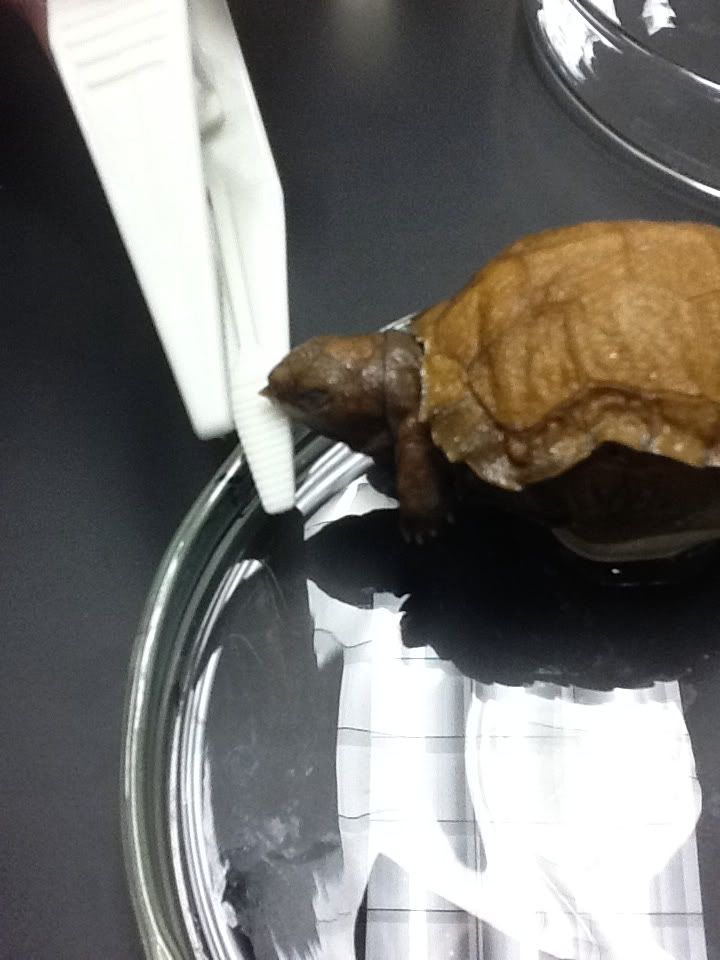
terrestrial adaption - this includes the yolk, an early food source, and other functional membrane layers. But how does the
hatchling emerge from this hard-shelled cage?! Duhh - an egg-tooth (seen above-right on the snout of this ageless young turtle)
Reptiles and early mammals both laid cleidoic
eggs (also called amniotic eggs); a chicken egg (as birds are highly derived reptiles) is an excellent example of an amniotic egg. Amniotic eggs are much more complex than anamniotic eggs (gelatinous amphibian
eggs that can rely on simple diffusion due to their aquatic environments). Amniotic eggs have crystalized calcium
carbonate shells that retain water while allowing diffusion of gasses through
small pores. Everything that the embryo needs
to survive early development can be found within the egg; an incredible
terrestrial adaption - this includes the yolk, an early food source, and other functional membrane layers. But how does the
hatchling emerge from this hard-shelled cage?! Duhh - an egg-tooth (seen above-right on the snout of this ageless young turtle)
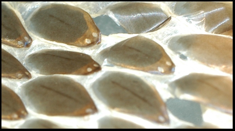
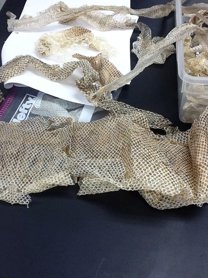
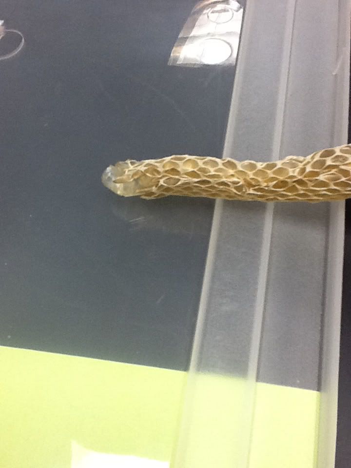
Reptilian skin has several layers, but is most often covered
by scales, or feathers (in the case of birds, which we don't really care about - lol). There are several different types of
scales and each type can be found in various places on any one specimen’s body. However, not all reptiles have scales, like our friend Zagnut, the soft-shelled turtle.

Here is an example of large rectangular scales on a cayman (right)
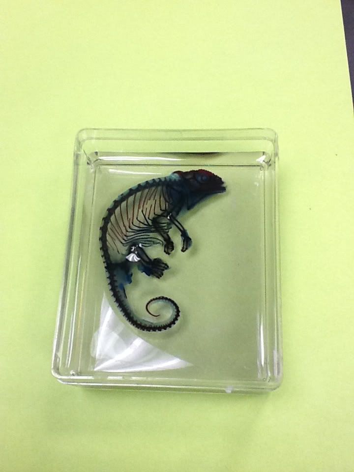
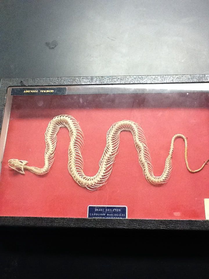
 The skeleton of tetrapods, in the case of most reptiles and mammals, evolved to endure a less aquatic life. This required beefing up the pelvic and pectoral girdles and strengthening the vertebral column (fusing many bones). Other notable adaptions include a more derived atlas-axis complex (pictured to the left), where the skull attaches to the start of the spine. This adaption allowed freer movement of the head for feeding. This is beneficial as pressure differences of air and water present different adaptational challenges.
 |
| This is a fine example of a tetrapod skeleton! (crocodilian) |

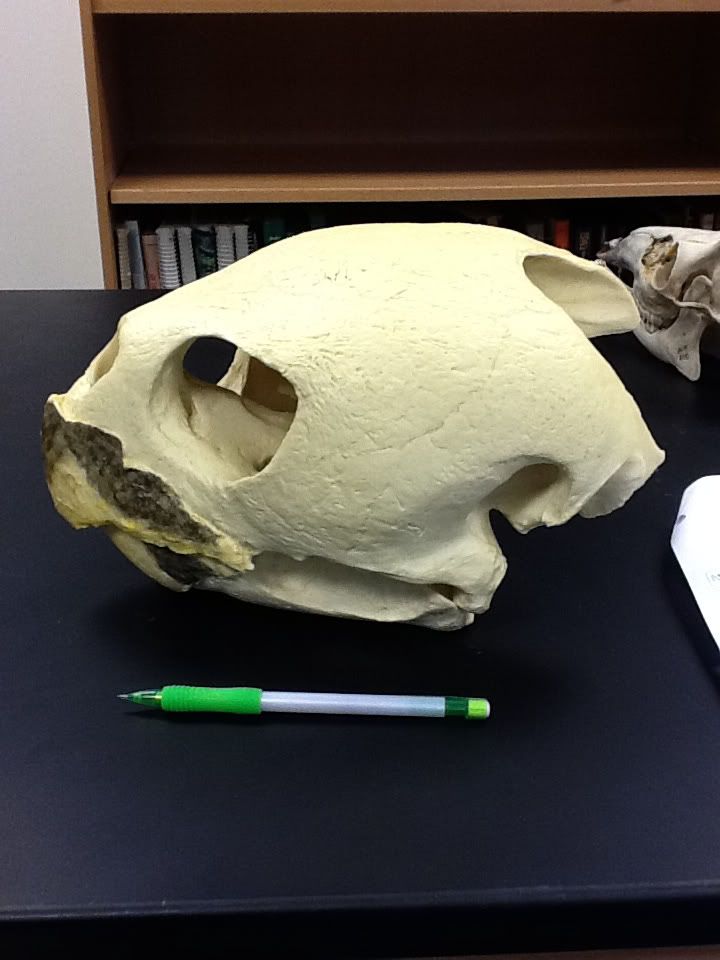
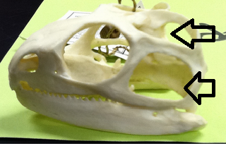
 The skulls of extant reptiles (those living today) are characterized as either diapsid skulls (snakes, lizards, crocodiles) or anapsid skulls (in the case of turtles). Reptile skulls may differ in the shapes of fused bones; however, the major differences lie in the presence or absence of fenestre - these are gaps or holes on the skull in certain places. Excluding the nose and eye holes, anapsid skulls completely lack fenestra (as did the skulls of the ancestral reptiles), while diapsid skulls exhibit both a supratemporal (just above and behind the eye socket) and infratemporal fenestre (just below and to the outside of the supratemporal)


 Cha-cha-cha!!
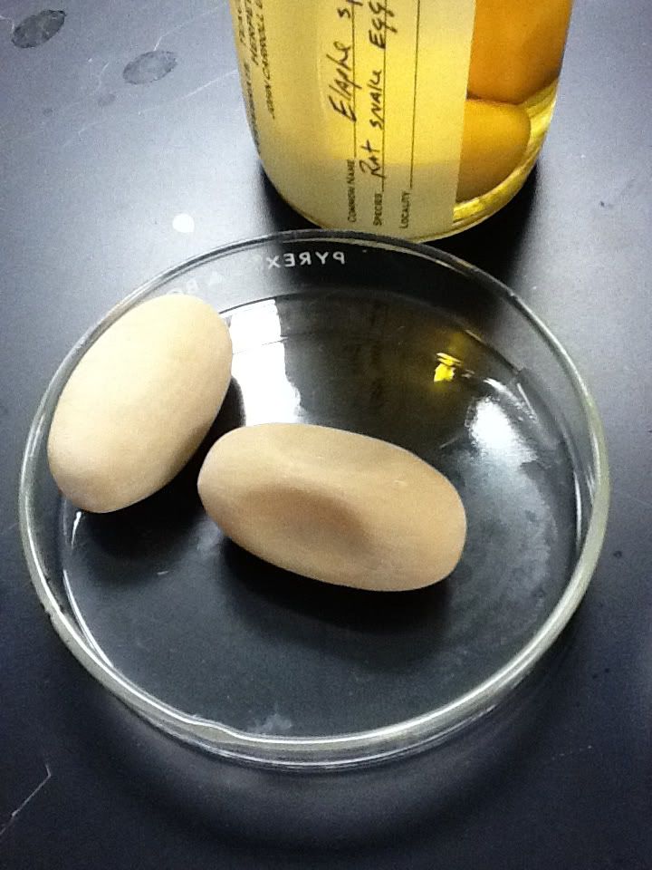


