When: Friday 20 January, 1:30-4:30pm
What We Did: Welcome to the first Reptiles lab of the semester! This week we looked at anatomy of many different reptiles, including snakes, turtles, lizards, and even chickens (yes, birds are reptiles!). Our lab is lucky enough to have various skeletons, eggs, and whole preserved reptiles to learn about different structures. Below is an overview of what we observed.
Eggs: Our lab had snake, snapping turtle, chicken, and ostrich eggs. The snake egg was smooth and leathery while the snapping turtle egg had the consistency of a ping-pong ball; i.e. it was perfectly round and very easy to squish. Not all turtle eggs are like ping-pong balls; ironically, softshell turtles have rigid egg shells.
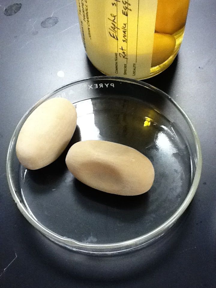
Snake egg (leathery, easily indented)

Ostrich egg (notice the small indentations, or pores, in the outer shell, used for the embryo's gas exchange). The shell's outer layer is made of shell units, composed of calcium carbonate, and all units are separated by these pores.
We then cracked open a regular chicken egg. After eating eggs for so many years, you would think we would know all there is to know about them, but we learned that eggs are pretty complex. If you have ever peeled a hard-boiled egg, you know that the shell contains 2 layers (outer & inner organic layer). However, the inside is not just yolk and "egg whites"; the albumin, or clear part of the internal egg, has multiple layers - external, middle, and internal albumin. The internal albumin is hard to detect, but if you crack open an egg in a dish, you can "peel" apart the external and middle albumin; the external has a liquid consistency while the middle is gelatinous.
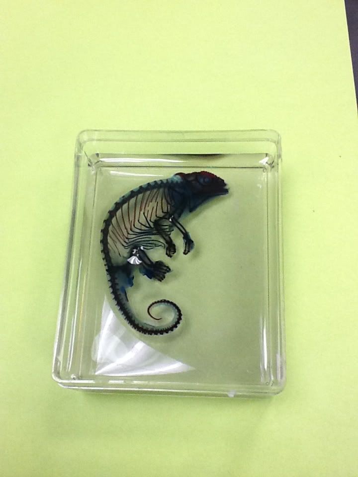
Embryos: We examined some preserved, double-stained turtle embryos. This means the body has been disintegrated down to cartilage and bone, both of which are stained different colors (cartilage is blue, bone is red). The embryos were small and hard to take a picture of, but below is an example of a chameleon that underwent the same staining process.
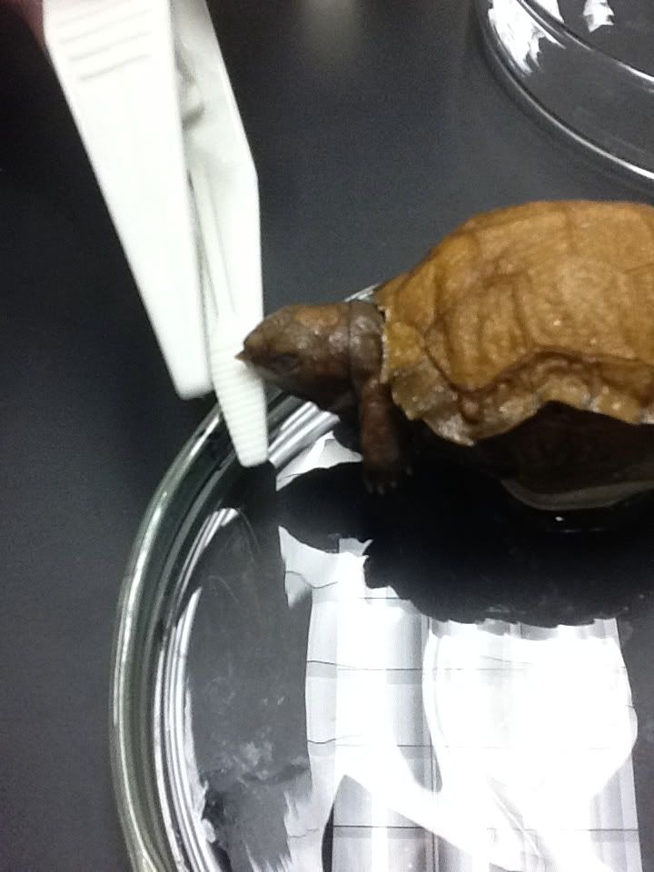
Hatching: Below is a turtle embryo with an egg tooth (protuberance between forceps). This tooth is used to break through the double-layered eggshell when the offspring is ready to emerge.
Skin: Even reptile skin is complex, with three distinct layers. When the reptile is ready to shed (called ecdysis), the "old" skin is the uppermost layer while new cells are created closer to the body. We had some really nice snake skins to analyze different sizes of scales.
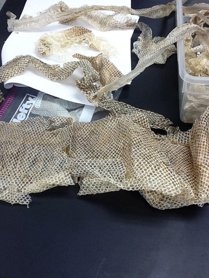
Multiple snake skins (largest is boa skin)
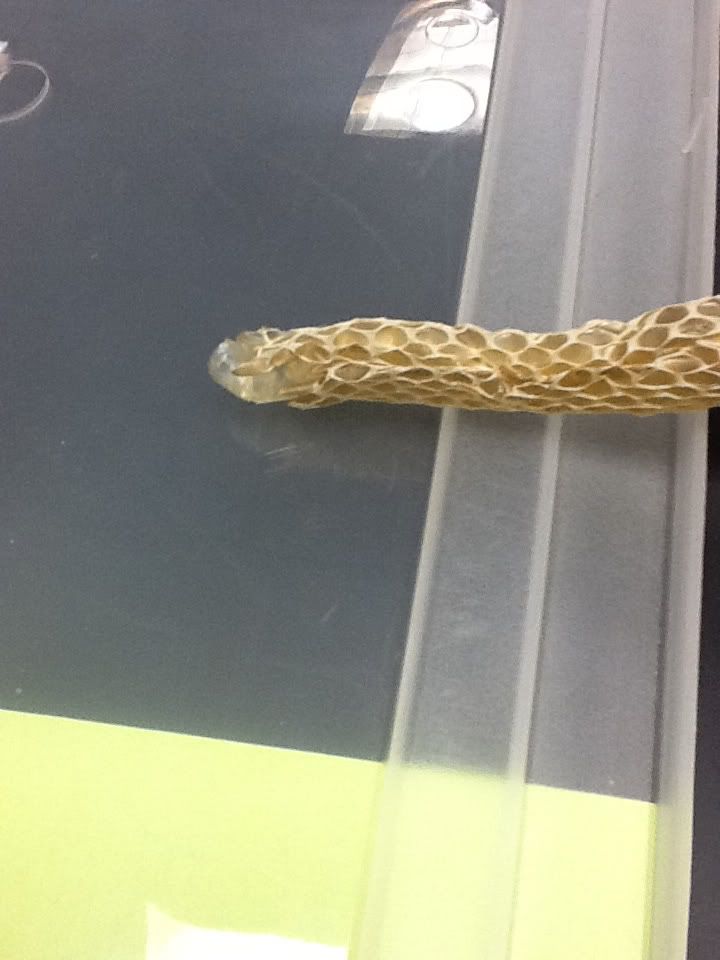
The edge of skin (left) shows the thin inner layer, while the remainder shows both layers of this snake's shed skin.
Scales: Reptiles have scales for thermoregulation (temperature), osmoregulation (water), and protection. Most scales are either rectangular or circular. However, some species can be distinguished by their combinations of shape and position; there exist overlapping circular (called cycloid), adjacent (called juxtaposed; there can be rectangular or circular juxtaposed), and mucronate (i.e., with a ridge down the scale's center) scale types. In addition, scales can be smooth or keeled (raised).
The 5-lined skink (Eumeces fasciatus) displays smooth cycloid scales. The cycloid scales completely line the body.

The gecko that was examined portrays a few different scale types over the body. The dorsal, or back, side of the body is composed of granular scales, which are raised and bump–like. There are rectangular scales on the tail while the ventral, or belly, side is composed of smooth juxtaposed scales.

Rectangular scales were examined on the green iguana (Iguana iguana), spiny tailed iguana (Ctenosaura similis), caiman (family Caimaninae), and the legless lizard (pictured below). The iguanas both have small smooth rectangular scales on their body, while the tail of the spiny-tailed iguana is keeled with larger rectangular scales. The scales of the caiman are large and tough, with some keeled around the tail. The legless lizard has small, keeled rectangular scales throughout its body.

Glands: This view of the gecko (below) shows the femoral (running along the femur, indicated by red circles) and precloacal (above the cloaca, or "all-purpose hole") pores. The femoral pores, more commonly found on males, are used to secrete hormones to mates or to mark territories.

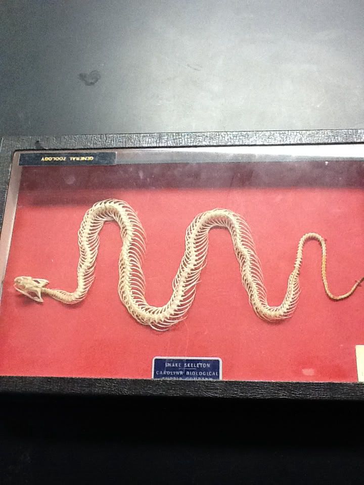
Skeletons: The anatomy of turtles is very unique. Turtles have very short limbs and a short tail. The vertebral column of a turtle is fused to its shell.

This snake skeleton demonstrates even more morphological variation from other reptiles. The skeleton does not contain appendages but is lined with ribs articulating the vertebrae.
Skulls: Reptiles display a variety of temporal fenestration, where fenestrae are large openings between bones in the temple. As a reference, our temples are between our eyes and ears, and can be felt when we open and close our jaws. There are evolutionary trends among fenestrae as modifications were made to the temporal regions of the skull to reduce weight and increase strength.
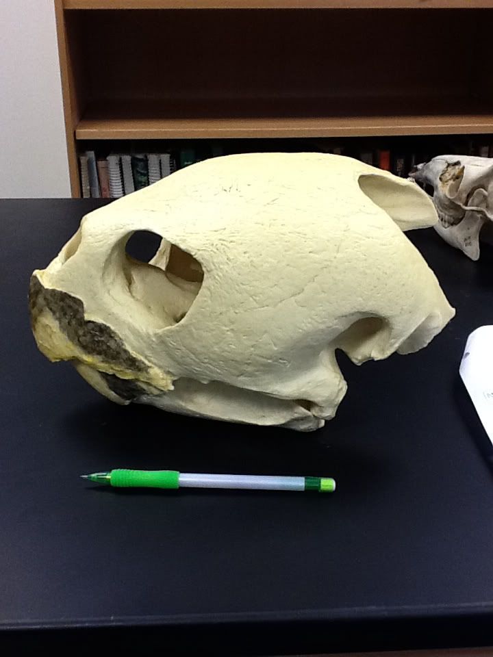
This turtle skull is anapsid. This is the most ancestral condition, with no temporal fenestrae (the large opening you see once held the organism's eye). The pencil gives you an idea of how big this skull is (it rivals a human's!).
Mammals have synapsid skulls, with only 1 hole posterior (behind) the orbital (eye) fenestra, clearly seen from the drawing below.

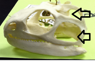
Below is a skull of a tuatara with 2 holes (fenestrae) posterior to the orbit, making it diapsid. The top arrow is pointing to the supratemporal fenestra while the bottom points to the subtemporal fenestra.
No comments:
Post a Comment