Monday, January 30, 2012

Reptiles lab 2 - Turtles (Thompson & Serna)

Where: JCU lab
When: Friday 27 Jan 2012; 1:30-4:30pm


What We Did/Background: This week was all about the Testudines, or turtles. These awesome creatures have been around for a LONG time - the first definitive turtle was Proganochelys from 210 million years ago! Today, 323 species of turtles are known to exist and compose 14 families. Turtles can be found in almost any habitat, on land or in water, but are not found on Antarctica and in the extreme artics. Overall, we looked at turtle anatomy, learned about diversity, and saw some live turtles!

ANATOMY - Skeleton
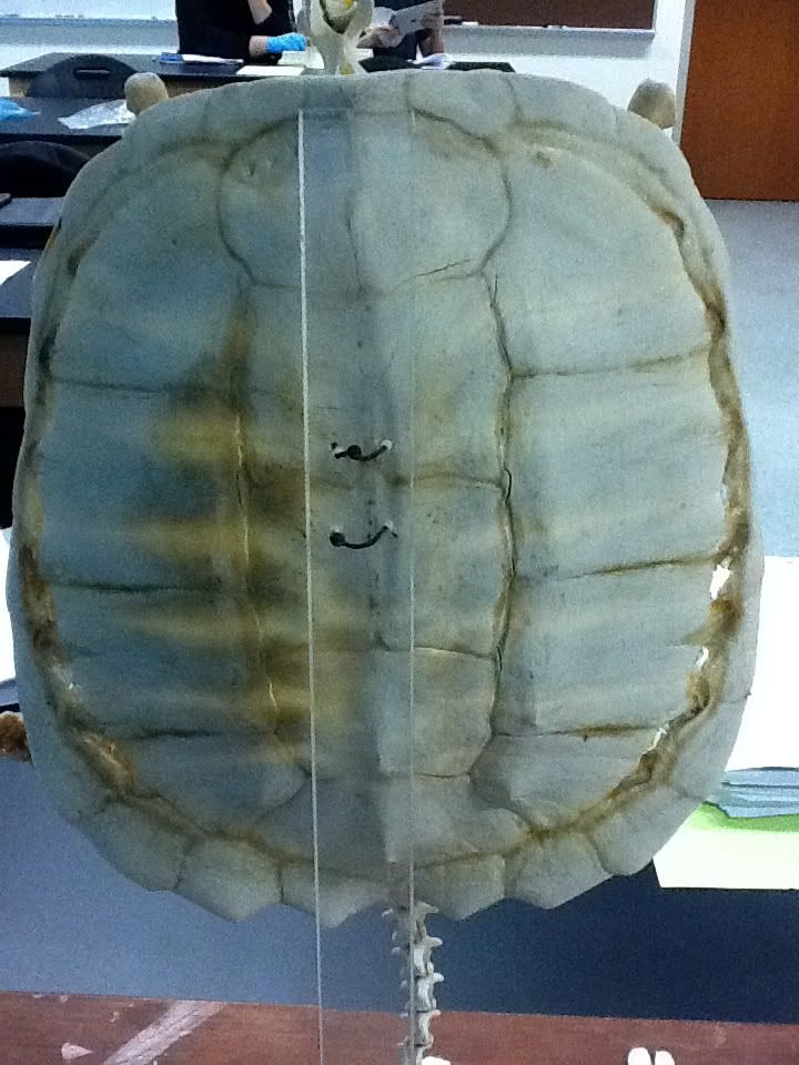
If you envision a turtle, chances are you see a head, limbs, and tail sticking out of a large shell. While most shells just look like big domes, they are actually pretty complex. The top (dorsal) shell is called a carapace while the bottom (ventral) is a plastron. Both the carapace and plastron are composed of bones and keratinous scales called scutes. If you look at the underside of the carapace, you can also see ribs fused with vertebrae.


Dermal bones


Plastron internal

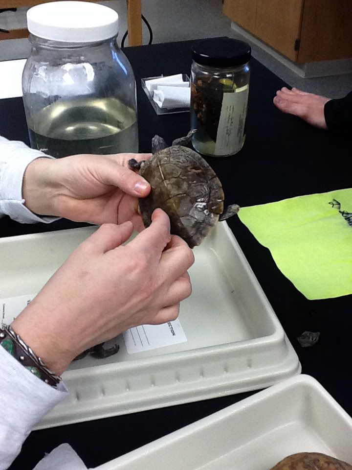
Like other reptiles, some turtles can seasonally shed both scales and scutes. One preserved specimen gave an example of scute loss (with the help of Dr. Sheil):
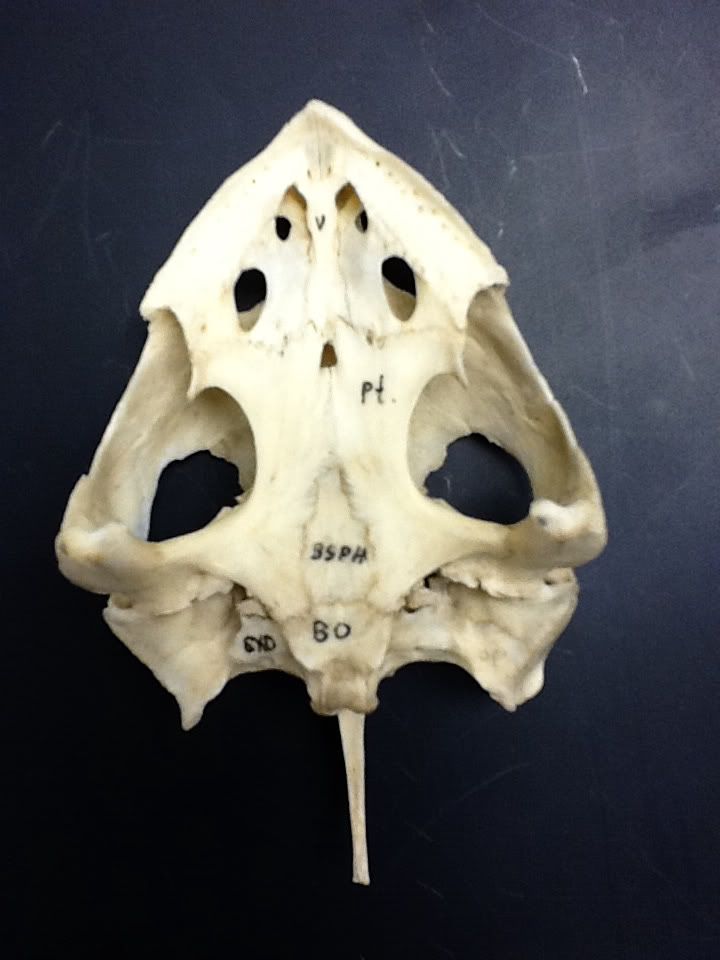


ANATOMY - Skulls
Turtles have anapsid skulls without temporal fenestrae, or holes behind the eyes (see the end of last week's blog). Below is a ventral, or underside, view of a turtle skull. The black markings indicate different bones - there are 10 here, just on the ventral side!


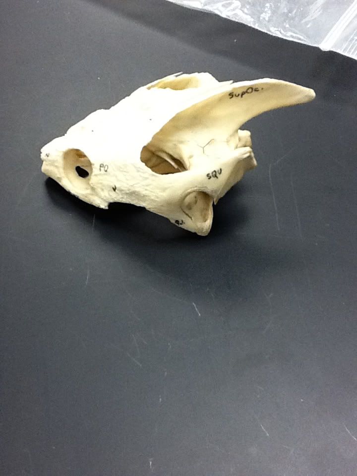
Here is a side (or lateral) view of a skull. Notice the big pointy bone towards the rear - this is the supraoccipital bone. This particular species emulates a temporal emargination, or a big gap between bones at the skull base where the supraoccipital bone is.

ANATOMY - Reproduction
All turtles are oviparous, meaning the mother does not house maturing embryos. The embryos develop inside a hard-shelled cleidoic egg (see last lab for more!). Male turtles have a copulatory organ and have slightly concave (or indented) shells to help position themselves on the female shell during copulation. The sex of a turtle can also be determined by gently pulling out the tail and noting the location of the cloaca. On males, the cloaca will be beyond the posterior end of the carapace, and the tails are more robust. Females have smaller tails and the cloaca does not pass the outer margin of the carapace.

Below is a picture of a male turtle. If you look at the posterior end, there's no need to tell you why this is a male.


DIVERSITY
Overall, turtles are divided into 2 groups: Pleurodira and Cryptodira. The main distinction between these two clades is that the Pleurodira move their heads to the side of the carapace as a defensive posture while the Cryptodira scrunch their head in an S-curve that draws the head directly into the carapace. Other traits that characterize these clades involve the “hips” and jaws. Pleurodirans have their pelvic girdles fused to the plastron while the cryptodirans have a flexible articulation (or joint) of the pelvic girdle with the plastron. The jaws of cryptodirans connect to the otic (ear/hearing) capsule while the jaws of pleurodirans are joined to the pterygoid (a bone on the ventral side of the upper half of the skull, essentially where our palate is). Recent molecular data also separates these clades.


A cryptodiran demonstrating its mastery at hide-and-seek.

In our lab, we had specimens of Cheloniidae (hard-shelled seaturtles), Chelidae (Australoamerican side-neck turtles), Chelydridae (snapping turtles), Emydidae (cooters, sliders, American box turtles), Kinosternidae (mud & musk turtles), Podocnemidae (Madagascan big-headed & American side-neck turtles), Testudinidae (tortoises), and Trionychidae (soft-shell turtles). Of these, only Chelidae and Podocnemidae are pleurodirans.

Cheloniidae (hard-shelled seaturtles) (Cryptodira)

These turtles range from the tropics to arctic and subarctic oceans. Their forelimbs act as flippers and are used in a figure-8 motion to swim. Cheloniids have amazing site fidelity, where mothers return within meters from where they were hatched. These sea turtles have a dietary range from seagrass to crustaceans to sponges, depending on the species. They are long-lived, reaching sexual maturity after 25 years.


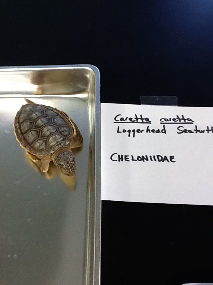
Caretta caretta - loggerhead sea turtle


Chelidae (Australoamerican side-neck turtles) (Pleurodira)

This family can only be found in the southern hemisphere, specifically in South America, Australia, and Indonesia. All have a flattened skull & shell and are primarily aquatic. Some species of Chelidae have extremely long necks. Some genera include Phrynops and Chelodina.

Chelodina spp.


Chelydridae (snapping turtles) (Cryptodira)

These turtles are naturally found in eastern North America, Mexico, Central America, and northern South America. One prominent feature of these turtles is their greatly reduced, cruciform-shaped plastron. Relative to the body, chelydrids have the longest tails. The 2 genera of this family are Chelydra (common snapping turtle) and Macrochelys (alligator snapping turtle). The common snapping turtle is native to Ohio while the alligator snapping turtle is only from the southern United States. Macrochelys has prominent ridges along its carapace, 3-4 supramarginal scutes, and a tongue lure used to attract prey while Chelydra has none of these characteristics.

Chelydra serpentina


Emydidae (cooters, sliders, American box turtles) (Cryptodira)

This family is broken up into 2 subfamilies: Emydinae and Deirochelinae. Emydinae consists of Calemys, Clemmys, Emys, Emydoidea, Glyptemys, and Terrapene. Deirochelinae contains the genera Chrysemys, Deirochelys, Graptemys, Malaclemmys, Pseudemys, and Trachemys. The family characteristics include a large plastron that is occasionally hinged. This allows the head and forelimbs to not only be protected but completely enclosed within the shell. These turtles exhibit sexual dimorphism, where the females can be twice as big as the males. As a whole, emydids compose the greatest Ohio diversity of turtles.

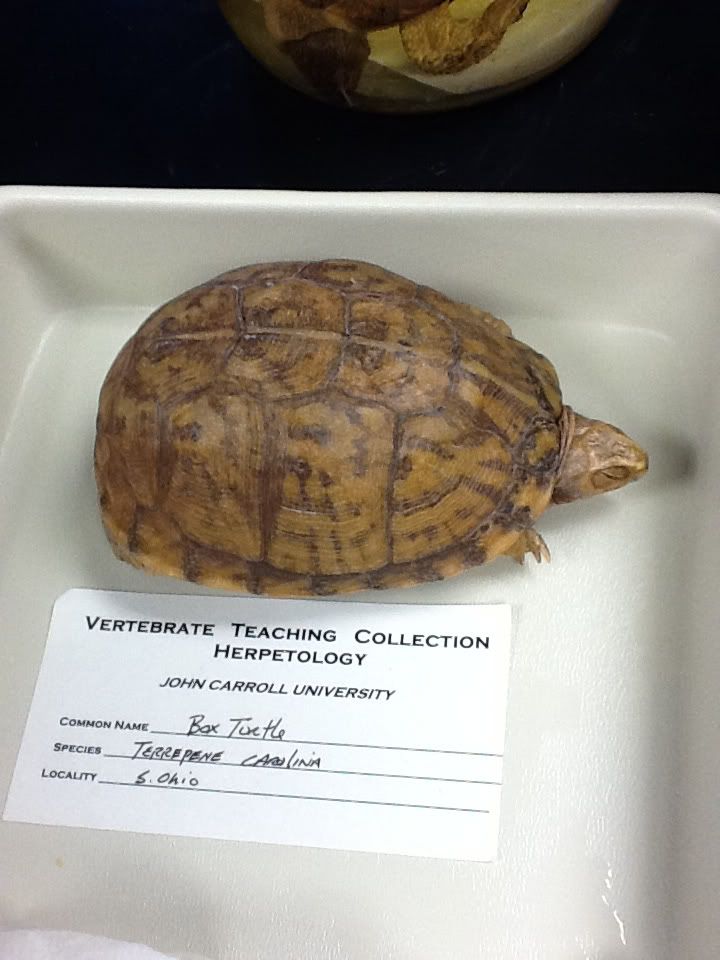
Box turtle - Terrepene carolina


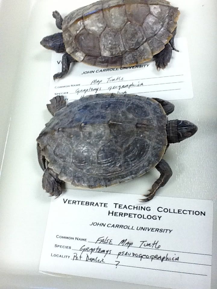
Graptemys geographica and G. pseudogeographica


Kinosternidae (mud and musk turtles) (Cryptodira)

This family of turtles is native to North, Central, and the northern part of South America. They are some of the smallest turtles and have oblong domed carapaces with less than 10 marginal scutes. While these turtles are aquatic and are commonly found walking on the bottom of aquatic surfaces, they hibernate on land.


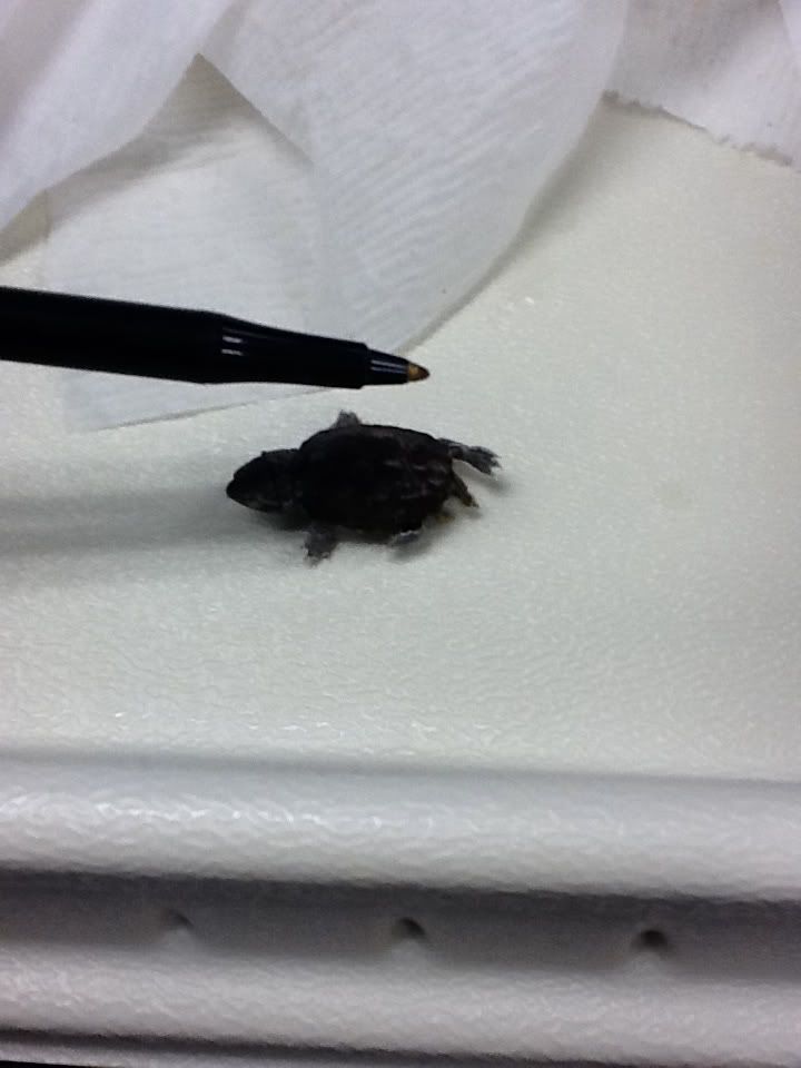
Sternotherus odoratus - stinkpot baby (so small!)


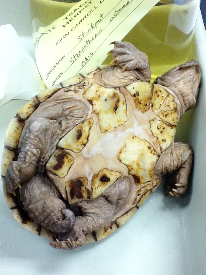
Stinkpot - notice the highly reduced plastron


Podocnemidae (Madagascan Big head turtles & American side neck turtles) (Pleurodira)

Podocnemidae is native to northern South America and Madagascar. These opportunistic carnivores and herbivores are found in rivers, marshes, and swamps. Podocnemids have large bodies and heads with powerful jaws.


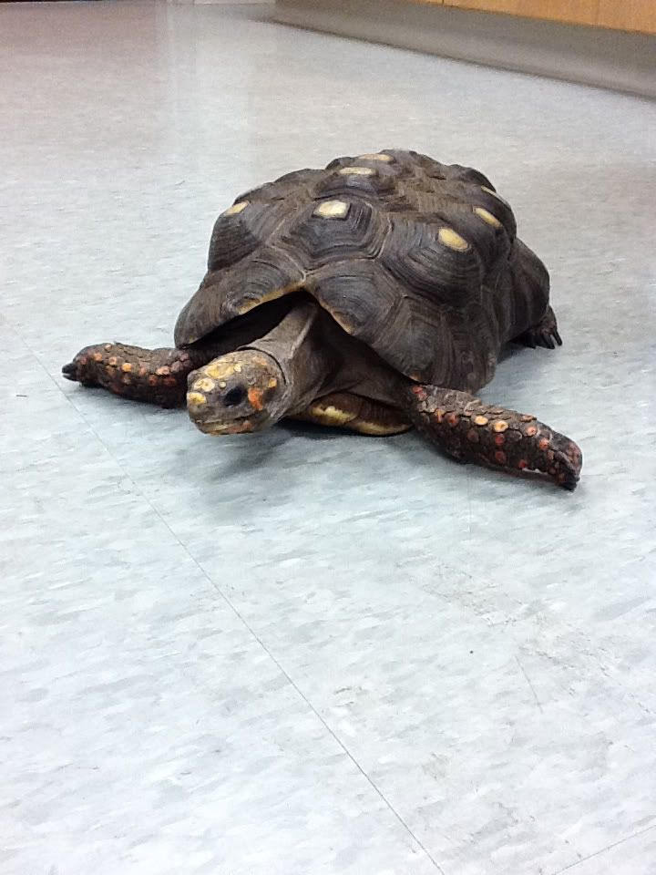
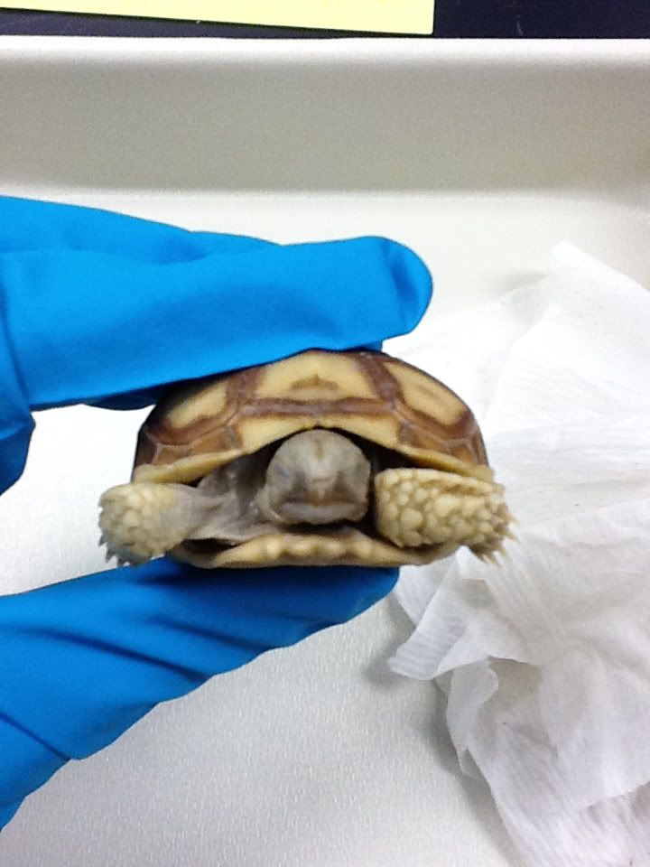
Testudinidae (tortoises) (Cryptodira)

The tortoises are large, terrestrial turtles that occupy North and South America, Europe, Asia, Indonesia, Galapagos, Seychelles, and Madagascar. Members of this clade have large, heavy, highly domed carapaces with large, armored scutes. They occupy grasslands, deserts, and other dry environments. These herbivores live to a very old age and have long stages of incubation and maturation.

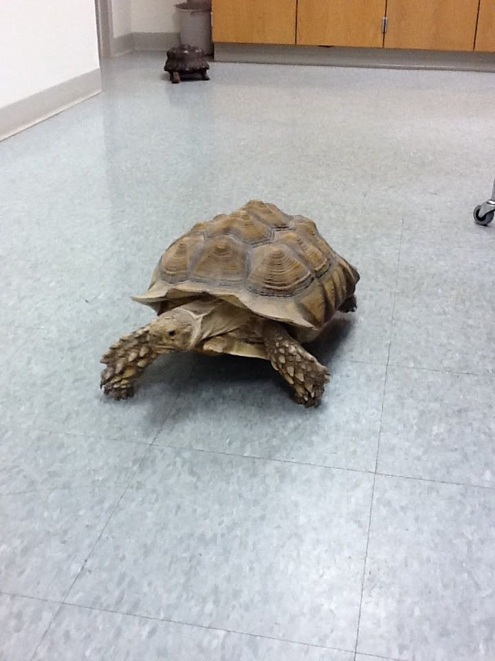
Geochelone sulcata, aka Samson the tortoise! No offense to the other turtles in our lab, but Samson was the highlight of the day.


Trionychidae (soft-shell turtles) (Cryptodira)

The soft shelled turtles are from North America, southeast Asia, New Guinea, and Africa. There are two subfamilies - Trionychinae and Cyclanorbinae. These turtles are flatter than most turtles and lack dermal scutes; their outer covering is thick and leathery, helping them cutaneously respire (or, essentially, breathe through their skin). These opportunistic omnivores have a long snout and are very efficient swimmers.


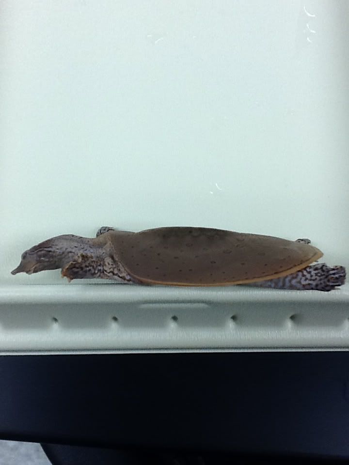
A lateral view of a softshell turtle. The lips that can be seen on the turtle help identify this family from its sister Carrettochelyidae.


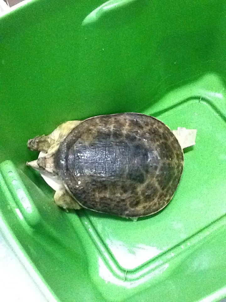
Apalone ferox, aka the soft-shelled Zagnut!


.
Ohio diversity!

Of the 300+ species of turtles, only 12 are native to Ohio:
Chelydra serpentina, Sternotherus odoratus, Apalone spinifera, A. mutica, Chrysemys picta marginata, Clemmys guttata, Emydoidea blandingii, Graptemys geographica, G. pseudogeographica, Pseudemys concinna, Terrapene carolina, and Trachemys scripta.
Chelydra is in Chelydridae, Sternotherus in Kinosternidae, Apalone in Trionychidae, and the rest in Emydidae.

Friday, January 27, 2012

Biology of the Reptilia Lab 1:
Authors: Matt Knestrick & Megan Thornhill


Purpose: We examined various features of amniotes (reptiles, specifically) to see what distinguished them from their evolutionary ancestors (fish and amphibians).


Eggs & Development
One of major features separating reptiles from their ancestors is the development of a cledoic egg. An egg is cledoic if it has developed a hard, calcium-carbonate outer shell, as opposed to a soft, membranous outer layer. This hard, outer shell is composed of shell units, which are small, teeth-like units that together make up the egg shell. This is essentially the difference between soft, gooey frog eggs, and the hard-shelled, Cledoic chicken eggs today.
Inside the eggshell there is an egg membrane, and an air sack, both of which are attached to the shell. Within the membrane is the outer, middle, and inner albumin, which are the gooey inners of the egg. In a chicken egg, these layers compose the egg white. Further inside the egg is the yellow yolk, and the yolk membrane that holds it together. Growing out of the yolk membrane is the chalaza. The chalaza are two membranous structures that anchor the yolk to the top and bottom of the egg .

Embryo development is also an important feature that separates amniotes from their predecessors. In amniotes, the embryos develop within the eggs into “miniature adults”. For example, a puppy looks like a miniature adult dog, and a baby human looks like a miniature grown-up. This is different from more ancestral animals, like amphibians, whose offspring first hatch as larvae, which exist on their own until they develop further into adults. The best example of this are frogs and toads, who offspring larval stage (tadpoles) look very different from the adults, and live very different lifestyles.

Skin & Glands
Reptile skin contains several different layers. The outermost layer is composed of dead cells this layer is called the stratum corneum, the next inner layer is composed of living cells of the deep epidermis, the innermost layer is the dermis, which contains nerves and blood vessels. One characteristic of reptiles is Ecdysis, or shedding. Reptiles will shed their outermost skin layers in order for growth of the organism.

Folding of the dermis and epidermis in these organisms produce specific irregularities which yield scales. Different combinations of these scale types can occur on the same organism. The types of scales that are expressed by each organism differ on different parts of the body. Some scale types include, smooth scales; granular scales, which appear bead-like and do not overlap; keeled scales, which have a distinguishable ridge on the scale; and scales with pits. Also, scales can be attached to the body in different ways, forming different characteristics. Cycloid scales overlap one another whereas juxtaposed scales do not overlap but simply sit close to one another.

Few glands are visible to the naked eye on most reptiles. Femoral glands are used for chemical communication between sexes as well as in marking territory. Usually only males possess these glands. If females do possess these glands then they are notably smaller than those on males of the same species.


Skeletal Structure
We also looked at reptilian skeleton to see the anatomical features that distinguish them from fishes.
The major skeletal feature that separates the amniotes from the fishes is the presence of skeletal girdles. These two girdles are known as the pectoral girdles (shoulders) that attach the forelimbs, and the pelvic girdle (hip-are) that attaches the hindlimbs. By having these girdles, the vertebral column was also noticeable sectioned off into the cervical, trunk, sacral, and caudelregions. These regions are used in almost all amniotes to describe body areas.
The skulls of amniotes are also different from ancetrals animals, in that they have a reduced number of skull bones Fish have a large number of skull bones, while amniotes have fewer. Amniotes’ skulls are also classified based on the number of finestra, or non-eye or nose holes, present. The ancestral condition, shared with fish, is anapsid, where that are no holes. Humans and some reptiles are synapsid, where only one hole is present. Some other reptiles are diapsid, where two holes are present. While technically, the term “apsid” refers to the number of arches found in the skull, this number is the same as the number of finestra (anapisids have no arches, diapsids have two), so the terminology works the same. These finestra act as channels for muscles to run through. In humans, these holes are underneath the check bone, and contain the muscle that operates the jaw.

Thursday, January 26, 2012

Laboratory #1: General Anatomy of Reptiles - Chris K & Cait F

by Cait Falasco and Chris Koch
Laboratory #1: General Anatomy of Reptiles

Who came first? The chicken or the egg?

In this case, we’re starting with the egg! (If you guessed chicken, unlucky!)
The beginning of the life of reptiles…these transitioning land pioneers needed to start their life in a warm, protective environment so nature’s selective pressure gave them the cleidoic egg.


The Cleidoic Eggs comes from the Greek kleistos meaning “closed” and these eggs, sometimes called “amniotic eggs”, are characteristic of amphibians and REPTILES! Assuming life begins at conception – controversial, we know - these developing little embryos live a very sheltered life in their beginning stages, simply because the egg has everything they need!

Protective force field? Check. That’s the outer layer “egg shell” made up of reinforced with calcium carbonate shell units. Some eggs are hard, like ostrich or bird eggs, and others are soft and leathery like the turtle eggs shown. The crystalline columns of shell units help the embryo conserve food and water and resist desiccation and other threats of the terrestrial environment, while dimple-like pores on the shell surface to help the babies exchange gases.

All-you-can-eat buffet? Check. The yolk and three layers of albumen are full of proteins for the embryos to chow down on throughout their stay. The yolk is the orange-yellow ball in the center, the external albumen is the runny liquid clear portion of the egg, the middle albumen is the runny clear substance within, and the inner albumen immediately surrounds the yolk.

Comfort/Stability? Check. If membranes were gold chains, this egg would be in a rap music video. Not only does the shell have stabilizing outer and inner shell membranes, but the yolk is surrounded by the vitelline membrane as well. There is the air cell “air bag” at one end of the egg and two white bungee cord-like yolk suspenders called chalazae for support within the center of the albumen.

Skin. The skin of reptiles is covered by scales OR in those trying to show off, highly derived scales: feathers!

The 3 layers of reptile skin are:
  1. The Stratum corneum (the outer layer of skin)
  2. Stratum germinativum (under the outer layer)
  3. Dermis (inner layer of skin)
The outer dead layer of skin (Stratum corneum) is what is shed through a process called ecdysis. The 2 remaining layers (Stratum germinativum and Dermis) contain living cells. The Stratum germinativum is composed of living cells of the deep epidermis and the Dermis forms a armor-like protective layer that can be found in some lizards, turtles and crocodilians.
We observed snake sheds to look at the outer layer of Stratum corneum.
The snake skins allowed us to see the outer layer of skin as well as look at the different scale types...speaking of which...
Scales: Reptiles have a wide range of different types of scales. Scale type can be used to identify specimens by classifying them with granular, rectangular, cycloid, juxtaposed or keeled scales. These different scale types can be small or large, smooth or keeled with a ridge in the middle of the scale, overlap or not overlap and some can have pits. It is important to note that an individual specimen can have several different types of scales on different parts of the body. In lab we looked at many different specimens and looked at the different types of scales.
Large rectangular scales can be seen in the ventral surface of this caiman specimen above.
Here to the left, this 5-lined skink has smooth cycloid scales.

Glands. Reptiles have a diversity of pores but only a few can be seen by the naked eye. In lab we observed femoral or precloacal pores. These glands are used for communication between sexes and marking territories.
In this lizard specimen femoral pores can be seen along the femur.

Lastly, SKELETONS:
Skeletal structure varies morphologically among reptiles but all demonstrate the general regions of the skeleton of a Tetrapod. Notice the skull and cervical region of the neck, esp. bones 1 and 2 (Axis-Atlas). After the neck lies the trunk (neck to the end of the hip bones) which is broken into 2 parts: the upper thoracic region (with ribs directly connected via gastralia) and the lower lumbar region without ribs.
Also much like the anatomy of humans, the reptile skeleton has a lot of bones with fancy names. The sacral region is composed of the hip structure withthe pelvic, ilium and ischium bones. The tail beyond the sacral region is the caudal region.


Many groups of reptiles are defined by their cranial structures thought explain certain evolutionary history. In particular, fenestrae and vacuities are very important structures for these ends. The number and position of fenestra, Latin for “window” or natural openings in the skull are helpful for categorizing amniotes as anapsid (0), synapsid (1 subtemporal), euryapsid (1 supratemporal), or diapsid (2).

That’s it for this week! We’ll leave you with an extremely mature and thought-provoking picture of one of us examining a specimen very thoroughly.