 |
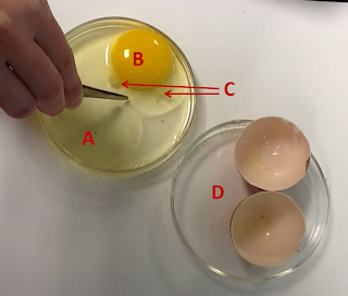
| Anatomy of a cleidoic egg: (A) albumin, (B) yolk, (C) chalaza, (D) shell |
The shell of a reptile egg is composed of two distinct layers: a hard, calcareous outer layer and a fibrous inner layer. Pores in the outer layer of the shell allow for gas exchange while the embryo is growing.
Inside the shell, we find the internal anatomy of the egg. In a fertilized egg, the embryo is encased in a nutrient-providing yolk; the yolk is anchored inside of the shell by the chalaza and is supported by the layers of the albumin ("egg white" in layman's terms).
 |
| Clockwise from bottom: snapping turtle, emu, ostrich |
While the internal anatomy of the cleidoic egg is similar among reptile species, the actual appearance of the egg varies greatly! Some, like birds, have a very hard shell, while others, like snakes, have a more flexible shell. Check out some of the different eggs to the left.
 |
| Egg tooth of late stage turtle embryo |
Reptiles exhibit direct development. This means that, when the embryo hatches from its egg, it looks like a mini-version of the adult. Many hatching reptiles have an adaptation called an egg tooth: this structure aids in breaking open the shell.
Skin, Scales, and Glands:
 |
| Dr. Sheil instructs the class on the different scale types |
The skin of a reptile is comprised of three distinct layers. The Stratum corneum is the layer of skin that is composed of dead skin cells. The second important layer, which is composed of living cells of the deep epidermis, is called the stratum germinativum. The last distinct layer discussed in lab is the dermis. The dermis forms the armor-like protective layer of some lizards, turtles, and some crocodilians.
Scales are important to pay attention to when attempting to distinguish specimens. Scale types range from smooth to keeled; some may overlap, and others may have pits. Different scale types can vary over the surface of individual bodies. Important scales to note are granular, small rectangular, large rectangular, cycloid, smooth juxtaposed, and keeled. Combinations of the types can occur. For example, some species may have keeled cycloid scales.
Glands of reptiles do not come close to the number amphibians have. Femoral glands are located on the inside of the upper hindlimbs and are visible to the naked eye, particularly in males. They manufacture chemicals that are used for communication and for marking territories.
 |
| Cleared and stained chameleon. Red is bone, and blue is cartilage |
When observing the skeleton of an alligator we see four distinct regions of the vertebral column. These include the cervical, dorsal, sacral, and caudal regions. An important anatomical feature of some reptiles is gastralia. Gastralia aid in the respiration of these particular reptiles.
Another important feature to point out in the skeleton is the skull. Reptiles can be classified as either anapsid or diapsid. These terms refer to the presence of (or lack of) a supratemporal fenestra and/or a subtemporal fenestra. Both are present in a diapsid skull, but none are present in an anapsid skull. Mammals have a synapsid skull, meaning that only the subtemporal fenestra is present.
 |
| Left to right: anapsid, synapsid, and diapsid skull conditions |
Learning about these characteristics helped us to further understand about what defines a reptile, particularly with regards to reptilian anatomy.
Stay tuned for our next adventure in BL 526 Biology of the REPTILIAAAAAA. Until next time...
KG & MH
 |
| Things get a little dangerous in the lab sometimes. |
I totally did the alligator eating my head pose too! (Cait)
ReplyDelete