Cleidoic Eggs and Development
First we looked at the cleidoic eggs of reptiles. We noticed how variable the thickness and texture of reptile eggs are, depending on the composition of the outer layer of the shell. One of the first characteristics we noticed was that the Rat Snake egg’s shell had a leathery texture.

The Snapping Turtle eggs resembled ping-pong balls and had a harder shell than the snake eggs. 
Next, we observed a broken Chicken egg shell and the internal anatomy of the egg. The outer layer of the
shell (1) is made of units of calcium carbonate, containing pores that allows for gas and water exchange. The inner organic layer (2) is made up of a fibrous layer and is easily seen after the egg has been broken.

After looking at the exterior components of the chicken egg, we looked at the internal anatomy of the egg.

The external albumin was much more runny than the more firm middle albumin. The chalaza is present on both sides of the yolk membrane and serves to anchor the yolk to the center of the egg shell. The air cell was visible on the inside of the empty egg shell and it looked like a bubble on one end of the egg
During development in the egg, many structures of the embryo become visible. The cleared and double-stained turtle embryo (below, on the left) is blue where cartilage has developed and the soft tissue has been cleared. It is easy to see the egg tooth on the late-stage turtle embryo (below, right) which is used to pierce through the membranes and egg shell. The yolk is still visible and attached to the ventral side of the turtle embryo, supplying it with nutrients while developing.


Integument: Skin, Scales and Glands
The skin of reptiles is composed of the Stratum corneum (the outer layer of dead cells), the Stratum germinativum (deep epidermis), and the dermis (contains nerves, blood vessels and osteoderms). The folding of the dermis and epidermis yields scales. There are many different types and arrangements of scales, which help herpetologists identify different species of reptiles.
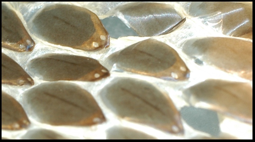
Below, are the scales of the Eastern Massasauga Rattlesnake (Sistrurus catenatus) which have a prominent ridge down the middle and are called keeled scales. These scales are shingle-like because they overlap.
 Keeled scales (Below, Left) are also visible on the shed skin of snakes (the shedding of skin is called ecdysis). We observed 2 apical scale pits on a shed (Below, Right).
Below (Left) is a picture of the cloacal plate, and the ventral and subcaudal scales of the S. catenatus. Venemous snakes generally have a complete cloacal plate and complete subcaudals. Brad and I noticed that a few of the subcaudal scales right beneath the cloacal plate were divided, but most of the scales were complete further down the tail. Below (Right) is a picture of S. catenatus' rattle (which is unique to genera Crotalus and Sistrurus), and by counting the sections, we can tell that this snake has gone through 8 sheds.


An example of a non-venomous Rat snake's divided cloacal plate and divided subcaudal scales:
 One of the specimens had a body similar to a snake because it didn't have legs, but it is actually a glass lizard (Ophisaurus). The features that give it away are the ear openings and the presence of eyelids, which are characteristic of lizards. We also noticed the glass lizard had a really long tail. The glass lizard has small rectangular scales on the dorsal half of it's body.

 To the left is a picture of
small rectangular, juxtaposed scales of an Iguana.

To the right is a picture of a gecko's femoral pores, which males develop when they are sexually mature, the glands secrete hormones.
Skeletal system
 To the right is a cleared and stained chameleon. This staining makes it easy to see where the skeletal system is (red) and where the cartilage is (blue). Below is a clear and stained Horned lizard. It is easy to see the vertebral column and regions of the skeleton in stained specimens.

Snakes have a large thoracic region (the region where ribs are present) and a smaller caudal region (absent of ribs).

To the left is an alligator skeleton, focusing on the Gastralia, where the ribs connect at the ventral midline. This whole picture shows the trunk region of the skeleton. The trunk region includes the lumbar region (lacking ribs) and the thoracic region (with ribs).

To the right are some of the different skulls of Amniota, and different modifications that either strengthen or lighten the skull with temporal fenestration, which are openings between bones.
1. Diapsid condition, common in reptiles (supratemporal fenestra and subtemporal fenestra)
2.Synapsid condition, often seen in mammalia (subtemporal fenestra)
3. Anapsid condition, without temporal fenestration. Seen in Testudines, Loggerhead turtle pictured here.
And now, a picture of Brad shaking some tail!!
 Cha-cha-cha!!
No comments:
Post a Comment